
In a recent study, immature and developing tapeworms were discovered in the spinal fluid of every MS subject tested. This post discusses the findings of this groundbreaking research and how it brings us closer to the cure for MS and other neurological diseases.
The cause of MS is unknown and there is no cure. I was diagnosed with multiple sclerosis 35 years ago and it is really sad that today, we are no closer to finding a cure for MS and we still don’t know what causes it.
In multiple sclerosis, the myelin surrounding nerve cells in the brain, spinal cord and eye becomes injured. Our standard of care assumes that the damage to myelin is caused by immune cells attacking myelin for some unknown reason. And as a result, all approved MS disease modifying drugs are designed to suppress immune cells in an attempt to reduce or prevent damage to nerve cells.
How MS is Diagnosed
The International Panel on Diagnosis of Multiple Sclerosis (Panel) is an international group MDs and PhDs that created the McDonald criteria which dictates how multiple sclerosis is diagnosed, how MS research should be conducted, and the regulatory approval of disease modifying MS drugs.
This panel has established that MS should be diagnosed through clinical observations and assessments, MRI imaging of the central nervous system for the presence of lesions, and when necessary, the examination of spinal fluid for the presence of antibodies.
Evidence That Multiple sclerosis is an infectious disease
Multiple sclerosis is an inflammatory disease. Inflammation is the immune system’s response to infection or parasites. Our immune system’s primary job is to defend us from disease-causing microbes (parasites). A parasite is any microbe that lives in us and causes us harm.
There are many different types of parasites that can infect humans. When they are present in the central nervous system (CNS), parasites can cause disease. Neurological disabilities can be caused by parasites like malaria, tapeworms, flukes, protists and soil transmitted worms.[i]
It is also well understood in science that many different parasites can cause lesions or cysts in the central nervous system.[ii][iii]
To review just a sample of peer reviewed studies showing the link between MS and various parasites CLICK HERE
Dr. Alan MacDonald is the first to report brain parasites in multiple sclerosis. In a previous study, he found many small immature roundworms in the central nervous system of every MS subject tested (see lecture here) and more recently, he also discovered immature and developing tapeworms in the central nervous system of MS patients.
In his 2021 study, “Novel Brain Compartment Tapeworm Larvae-Possible Environmental Initiators of Demyelination in Multiple Sclerosis,” Dr MacDonald examined the spinal fluid of 10 MS patients that was kept in a brain bank for research purposes. The dates of autopsies of the subjects were between 1984 – 2014.
All brain tissue from these subjects received a proper neuropathological examination to confirm the type of MS that the patient had and ensure there were no additional brain diseases or medical conditions in addition to MS.
“Thawed cerebrospinal fluid from ten patients was, for the first time, examined in this time capsule research study. All frozen fluids had been untouched since initial autopsy harvest.
The justification for the permanent omission of pathologist examinations of autopsy cerebrospinal fluids was the declaration by international expert panels in multiple sclerosis practice, that microscopy of multiple sclerosis cerebrospinal fluids is always without any value, either antemortem of post-mortem.”[iv]
If this panel understands that:
- the immune system is involved and active in multiple sclerosis (because of the presence of lesions and antibodies on the CNS)
- the immune system protects/defends us from infections /parasites
- it is well understood in the scientific literature that parasites in the central nervous system cause lesions and cysts
Then why would this Panel declare that it’s “of no value” for pathologists to use microscopy to search for possible parasites that may cause MS, especially when it is known that parasites cause lesions in the CNS and the cause of multiple sclerosis is unknown. Why are they content with not knowing what is causing the lesions in MS?
The Results of Dr. MacDonald’s Study
He found tapeworm oncospheres (hatched eggs) in two subjects and larval tapeworms in the cerebrospinal fluids of all 10 subjects tested. Multiple stages of growth of the cystic tapeworm larvae were found in all subjects.
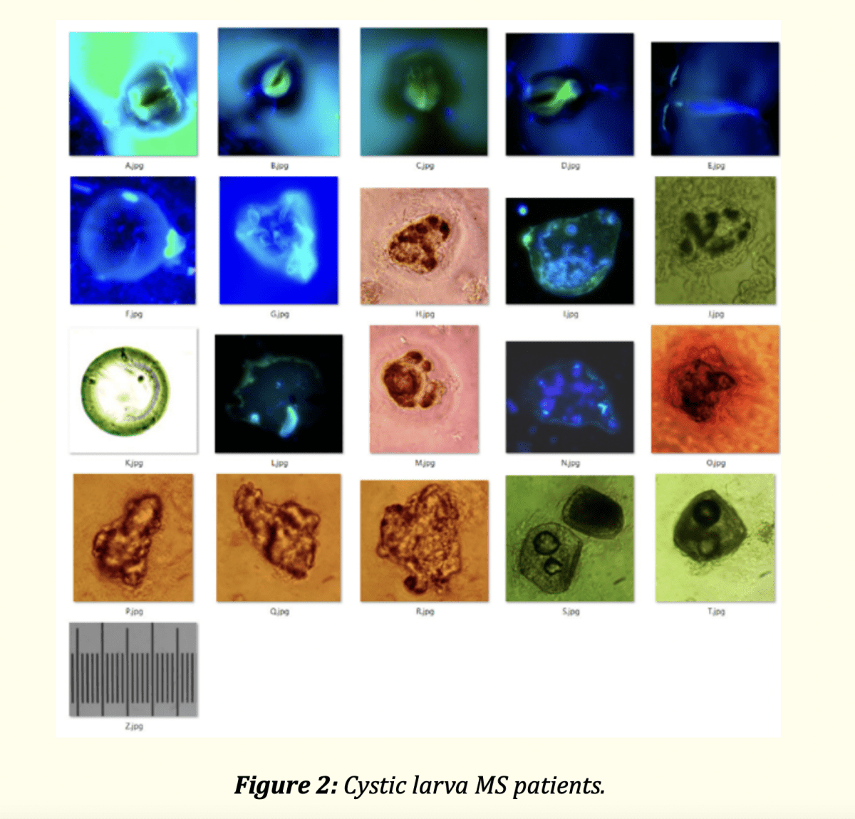
Image from Dr Alan MacDonald’s study
Juvenile tapeworms were found in two of the ten subjects.
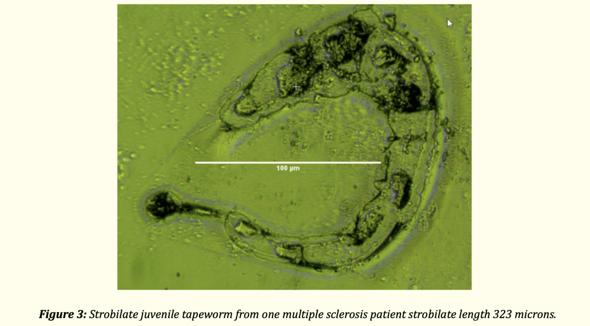
Image from Dr Alan MacDonald’s study
Dr. MacDonald observed various structures associated with the cystic larval form such as strips of tegument (a protective barrier), racemose membrane and corpuscles.
“Cestode hooklets were found in all ten patients and each patient demonstrated unique hooklets unlike those of any other patient in this group of ten, and unlike any hooklets of Taenia species or of echinococcal (tiny tapeworms) species of tapeworms.”[v]

Image from Dr Alan MacDonald’s study
No tapeworm eggs were identified in any patient.
He also observed that the tapeworm larvae were present in a bladder like membrane or sack (Coenurus pattern), where there were at least two and up to eight immature larvae inside the membrane of each cyst.
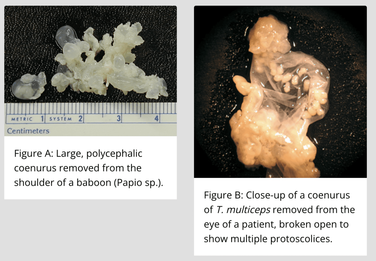
The dimensions of the parasites in the spinal fluid of all 10 MS patients were considerably smaller than the smallest known human tapeworm (Echinococcus species) and much smaller than cysts from the pork tapeworm.
He mentioned that there is a more recently discovered human brain and tissue tapeworm species (Versteria sp.) that when present in animals, the cysts, hooklets and developing tapeworms are minute and similar in size to what he found in the MS subjects. He did comment that there were likely multiple different species of tapeworm in the 10 MS subjects because the larvae varied in structure. He did not have enough DNA to identify the type of tapeworm present in the subjects.
In every case, these parasites were invisible to the naked eye of a neuropathologist and were not detected during the brain autopsy.
Dr. MacDonald’s findings are very significant because in animal cases (sheep, goats and other primates), it has been shown that brain demyelination occurs next to or beside the bladder/sack cysts of tapeworm larvae. He concluded that animal models help us to understand how these cysts in the CNS of humans could cause demyelination in the white brain matter of the 10 MS subjects. His research is the first to report brain parasites in multiple sclerosis.
Another Study Finds Cystic Structures in the Spinal Fluid of MS Patients
The aim of the study, Association between Multiple Sclerosis and Cystic Structures in Cerebrospinal Fluid was to search for infectious agents in the spinal fluid of MS patients.[vi]
The spinal fluid from ten patients with relapsing remitting MS and five controls without MS were examined by microscopy. Cystic structures were observed in spinal fluid of all ten MS patients and one of five control subjects who suffered from erythema migrans had cysts in the spinal fluid.
Neurocysticercosis
According to the CDC, neurocysticercosis is a preventable parasitic infection caused by larval cysts of tapeworms that infect the central nervous system and cause significant illness, disability and in severe cases death. It is considered a neglected parasitic infection because most health care providers lack the skill to identify or treat this condition.[vii]
Neurocysticercosis is the most common neurological infection cause by worms and is a major public health problem in most of the world. It is estimated that millions of people are infected with these cysts and only some become symptomatic during their life.[viii]
Symptoms of neurocysticercosis:
Neurocysticercosis disease can vary in severity from a completely asymptomatic infection to severe disease and death. This is determined by the number, size, and location of cysts in the CNS and intensity of the immune response. Cysts can survive for years or even decades in the CNS where intermittent neurological symptoms may be experienced. Neurocysticercosis mimics almost any neurological disorder.[ix]
Symptoms of neurocysticercosis include (but are not limited to):
- Headache
- Dizziness
- Poor balance
- Poor coordination
- Difficulty walking and falls
- Nerve pain in the spine or limbs
- Muscle weakness
- Muscle twitches or spasms
- Muscle paralysis
- Seizures
- Changes in thinking or behaviors – confusion or loss of consciousness for even a moment
- Tinnitus, hearing loss
- Nystagmus – involuntary, rapid and repetitive movement of the eyes
- Facial nerve palsy
- Trigeminal neuralgia
- Radicular pain – pain that radiates from the back and hip into the legs through the spine
- Impaired cognitive function
- Loss of taste or smell
Testing for neurocysticercosis:
According to the CDC, diagnosing neurocysticercosis usually requires MRI or CT brain scans. Blood tests may be helpful, but they may give a false negative test result in light of infections. If diagnosed with cysticercosis (tapeworm larval cysts), it’s important to also be tested for intestinal tapeworm infections.[x]
The metagenomic test can be used to analyze all of the DNA and RNA found in a sample of cerebrospinal fluid.
Treating neurocysticercosis:
When treating parasites in the central nervous system, it’s very important to be closely monitored by a healthcare professional to prevent significant or even life-threatening die off reactions.
Neurocysticercosis treatments may include:
- Anti-inflammatory treatments – anti-seizure medication and or steroids such as dexamethasone or prednisone
- Parasite medications – combined albendazole and praziquantel therapy may be more effective than albendazole alone[xi][xii][xiii]
- Minimally invasive neurosurgery.
It’s extremely odd that the Panel responsible for setting the guidelines for diagnosis and treatment of multiple sclerosis feels that there is no value in studying MS spinal fluid for the presence of infections when common sense and overwhelming evidence points to the fact that MS is an infectious disease.
Towards the bottom of page 170 of the position paper Diagnosis of multiple sclerosis: 2017 revisions of the McDonald criteria, the authors list the “Declaration of Interests” which reveals that many pharmaceutical companies and the Multiple Sclerosis Society have been generously financially supporting the Panel with grants, speaking fees, reimbursement for travel expenses and much more.
Is it not a conflict of interest when companies who make billions of dollars on the sale of their products, financially fund the Panel who produce the guidelines on how MS is diagnosed and which drug treatments are used? Is this why today we have many very expensive disease maintenance drugs and yet we are still no closer to a cure for MS?
There are real solutions to recover from parasites today!
To restore health, we must focus on treating the cause of inflammation, which are parasites. First, identify the enemy (parasites), then support the body and treat the parasites while following a holistic approach. When parasitic infections are treated effectively, we can overcome inflammation or disease.
If you’re frustrated with the fact that our standard of care STILL doesn’t offer a real solution for treating MS and other diseases, then click on the link below to watch Pam Bartha’s free masterclass training and discover REAL solutions that have allowed Pam and many others to live free from MS and other diseases.
CLICK Here to watch Pam’s masterclass training
References:
[i] https://www.ejmanager.com/mnstemps/51/51-1515268583.pdf?t=1677096864
[ii][ii] https://www.semanticscholar.org/paper/Parasitic-infections-of-the-spine%3A-case-series-and-Majmundar-Patel/efa6df10da344b402aa298c0c80daa8e488f7b6e
[iii] https://jesp.journals.ekb.eg/article_90704_51e0a7ae8ad24f9084e5d82006719829.pdf
[iv] https://www.ilads.org/wp-content/uploads/2021/05/ECMI-17-01109-Novel-brain-Compartment-Tapeworm-Larvae-in-Multiple-Sclerosis-10-patients-PDF.pdf
[v] https://www.ilads.org/wp-content/uploads/2021/05/ECMI-17-01109-Novel-brain-Compartment-Tapeworm-Larvae-in-Multiple-Sclerosis-10-patients-PDF.pdf
[vi] https://www.researchgate.net/publication/11570331_Association_between_Multiple_Sclerosis_and_Cystic_Structures_in_Cerebrospinal_Fluid
[vii] https://www.cdc.gov/parasites/resources/pdf/npis_in_us_neurocysticercosis.pdf
[viii] https://www.ncbi.nlm.nih.gov/pmc/articles/PMC6108081/#R38
[ix] https://www.ncbi.nlm.nih.gov/pmc/articles/PMC6108081/#R38
[x] https://www.cdc.gov/parasites/cysticercosis/diagnosis.html#:~:text=The%20diagnosis%20of%20neurocysticercosis%20usually,tested%20for%20intestinal%20tapeworm%20infection.
[xi] https://academic.oup.com/cid/article/62/11/1375/1745120?login=false
[xii] https://www.ncbi.nlm.nih.gov/pmc/articles/PMC6143370/
[xiii] https://www.ncbi.nlm.nih.gov/pmc/articles/PMC4926779/

Clinically diagnosed with multiple sclerosis at the age of 28, Pam chose an alternative approach to recovery. Now decades later and still symptom free, she coaches others on how to treat the root cause of chronic disease, using a holistic approach. She can teach you how, too.
Pam is the author of Become a Wellness Champion and founder of Live Disease Free. She is a wellness expert, coach and speaker.
The Live Disease Free Academy has helped hundreds of Wellness Champions in over 15 countries take charge of their health and experience profound improvements in their life.