Intestinal flukes are more common than we realize. They are large, flat parasitic worms that live in the small intestine and can cause digestive issues, anemia, toxemia, edema, allergic reactions, neurological symptoms and other symptoms of inflammation. This post discusses the symptoms of intestinal flukes, what they look like, how we become infected with them and how we can successfully treat them.
Flukes are oval or elongated trematode parasitic flatworms that infect humans. Different species of flukes infect different parts of the body. Blood flukes infect the blood vessels of the digestive or urinary tract. Intestinal flukes infect the lining of the intestines. Liver flukes infect the liver and bile ducts, and lung flukes infect the lungs.
Although there are thousands of species of flukes, about 70 are known to live in the human digestive tract. They are found worldwide but are most prevalent in East and Southeast Asia. It’s estimated that 40-50 million people are infected with intestinal flukes, mostly through contaminated food.[i]
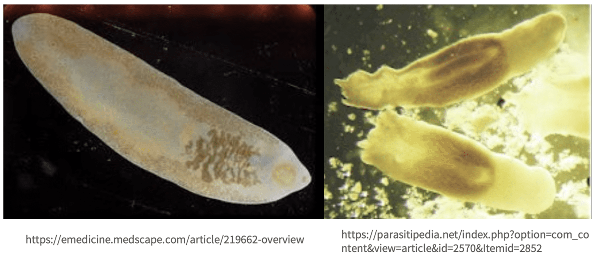
The most common human intestinal fluke is known as the giant intestinal fluke because it’s the largest fluke that infects humans and can grow to approximately 2 – 10 cm in length and 0.8 – 3 cm in width.[ii] It is mostly present in the intestines of pigs. Adult giant intestinal flukes live approximately 1 year.[iii]
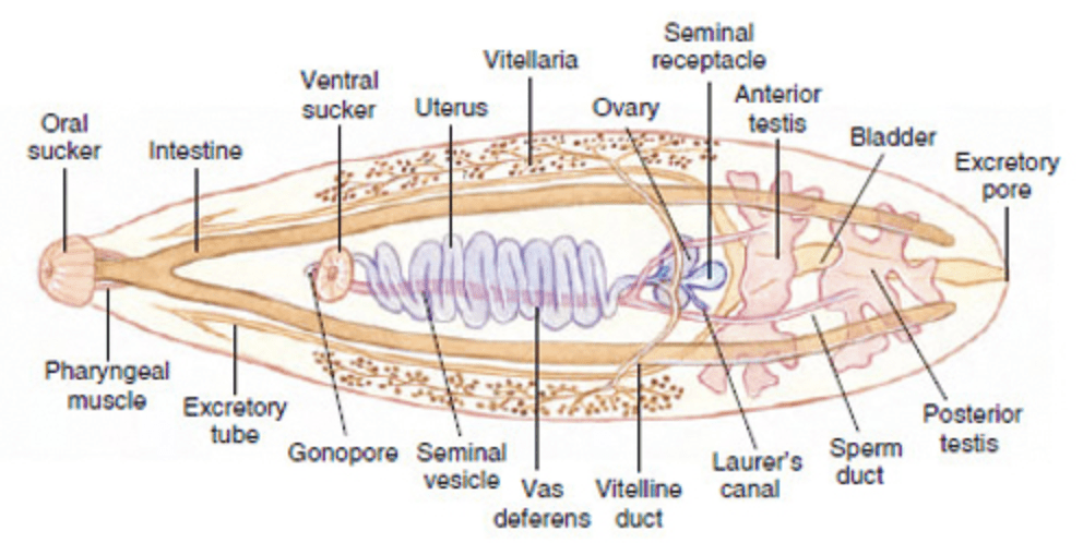
Image: https://www.bio.miami.edu/dana/330/330F19_15.html
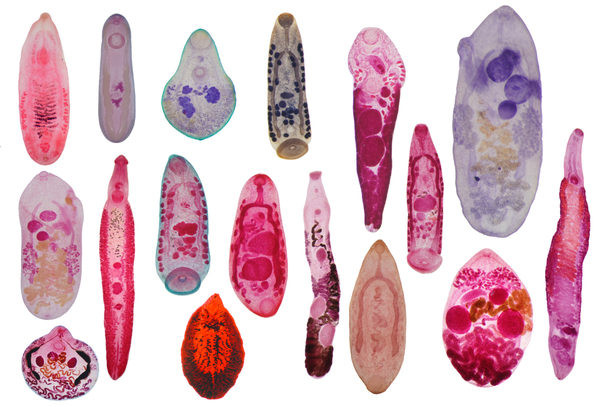
Images of Intestinal Flukes
How we become infected with intestinal flukes
Humans become infected through contaminated water, eating raw or undercooked aquatic plants, fish (salmon, trout, steelhead, shrimp, oysters, freshwater fish, snails) or meat (pork, poultry, beef) that is infected with the encysted larval form of the fluke.
After intestinal fluke larvae are ingested, they move to and live in the small intestine where they develop into adult flukes and attach to the intestinal wall in about 3 months. Female flukes produce eggs that are excreted in the stool.
Symptoms of intestinal flukes
In the early stages of infection, when small numbers of flukes are present in the small intestine, a person may have no or minimal symptoms. Hunger pangs can be an early symptom. One or two months after becoming infected with intestinal flukes, abdominal pain and diarrhea or constipation can occur.
Heavy intestinal fluke infections cause inflammation, ulcers and mucus where the flukes attach to the lining of the intestinal wall.[iv] Other symptoms include malabsorption, nausea, vomiting, weight loss, malaise, fever and intestinal obstruction. Allergic reactions and swelling of the face and legs can also occur and anemia may be present.[v]
Severe infections may also result in low albumin, vitamin B-12 deficiency and eosinophilia.
In severe infections, diarrhea may alternate with constipation. Some may experience bloody diarrhea.
As the worm burden increases over time, systemic inflammation and intoxication from metabolites produced by the flukes can cause:
- toxic and allergic symptoms such as edema of the face, abdominal wall, and lower limbs
- abdominal distention and pain
- anorexia
- nausea
- vomiting
- if diarrhea persists it becomes greenish-yellow with an exceptionally foul odor
- intestinal obstruction
- ascites – a build up of fluid in the space between the lining of the abdomen and abdominal organs
- some species of intestinal fluke larvae can cause myocarditis, heart failure, and brain bleeds
- intestinal fluke eggs in the spinal cord can impact motor and sensory function depending a lesion is located
- neurological symptoms
Severe intestinal fluke infestations can be fatal.
How are intestinal flukes diagnosed?
The diagnosis is made by finding the flukes or their eggs in stool or in vomit. Other tests include Merthiolate, iodine, formalin method or PCR.[vi]
Because stool tests are inaccurate and lead to false negative test results, energy testing is be helpful. Skilled healthcare professionals can use energy testing to determine which treatments test best for a patient.
CLICK HERE to learn more about the best parasite tests.
Treatment
Praziquantel is the drug of choice for most intestinal fluke infections, although niclosamide has also been shown to be a helpful treatment.
Artesunate has been used to treat certain types of intestinal fluke infections, with promising results.[vii]
Combination parasite drug protocols may be more helpful to eradicate persistent infections.
Tetrachloroethylene as capable of reducing faecal egg counts by up to 99%. Other parasite drugs that may be helpful include thiabendazole, mebendazole, levamisole and pyrantel pamoate, oxyclozanide, hexachlorophene and nitroxynil.[viii]
It’s very important to prepare before treating parasites by following the prep phase of the Live Disease Free program and to work with a practitioner who will prescribe the most helpful treatments at safe and effective doses.
CLICK HERE to learn watch Pam Bartha’s masterclass training.
What intestinal flukes feed on
Intestinal flukes feed on the cells and cell fragments of the host and digested food in the small intestine.
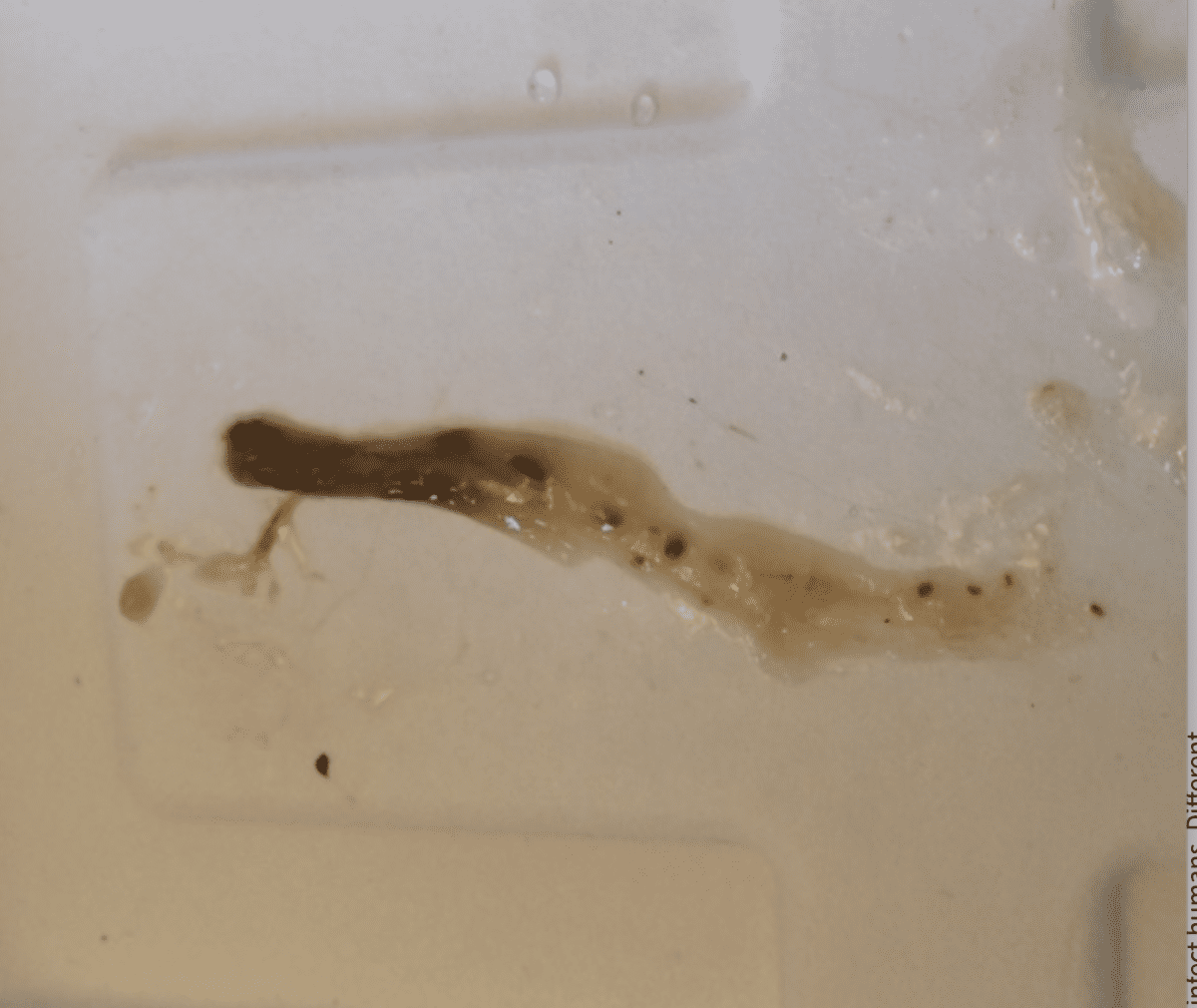
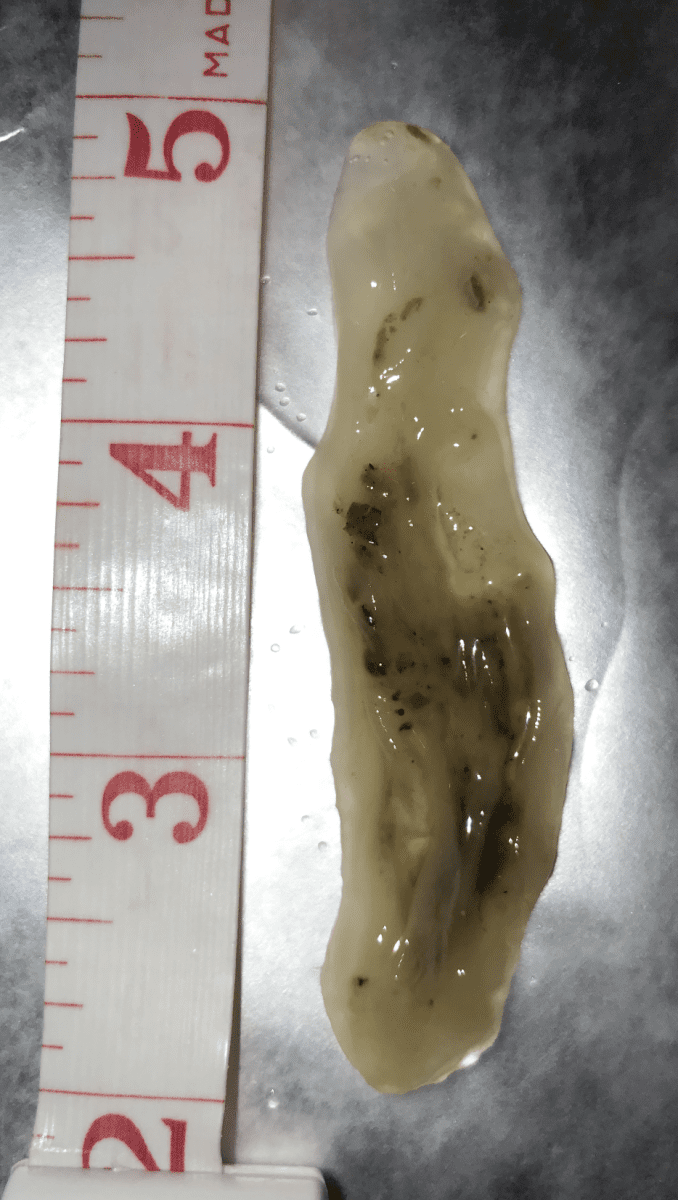
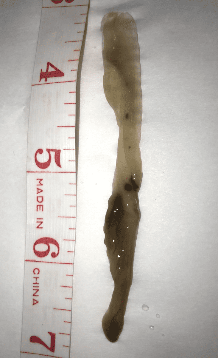
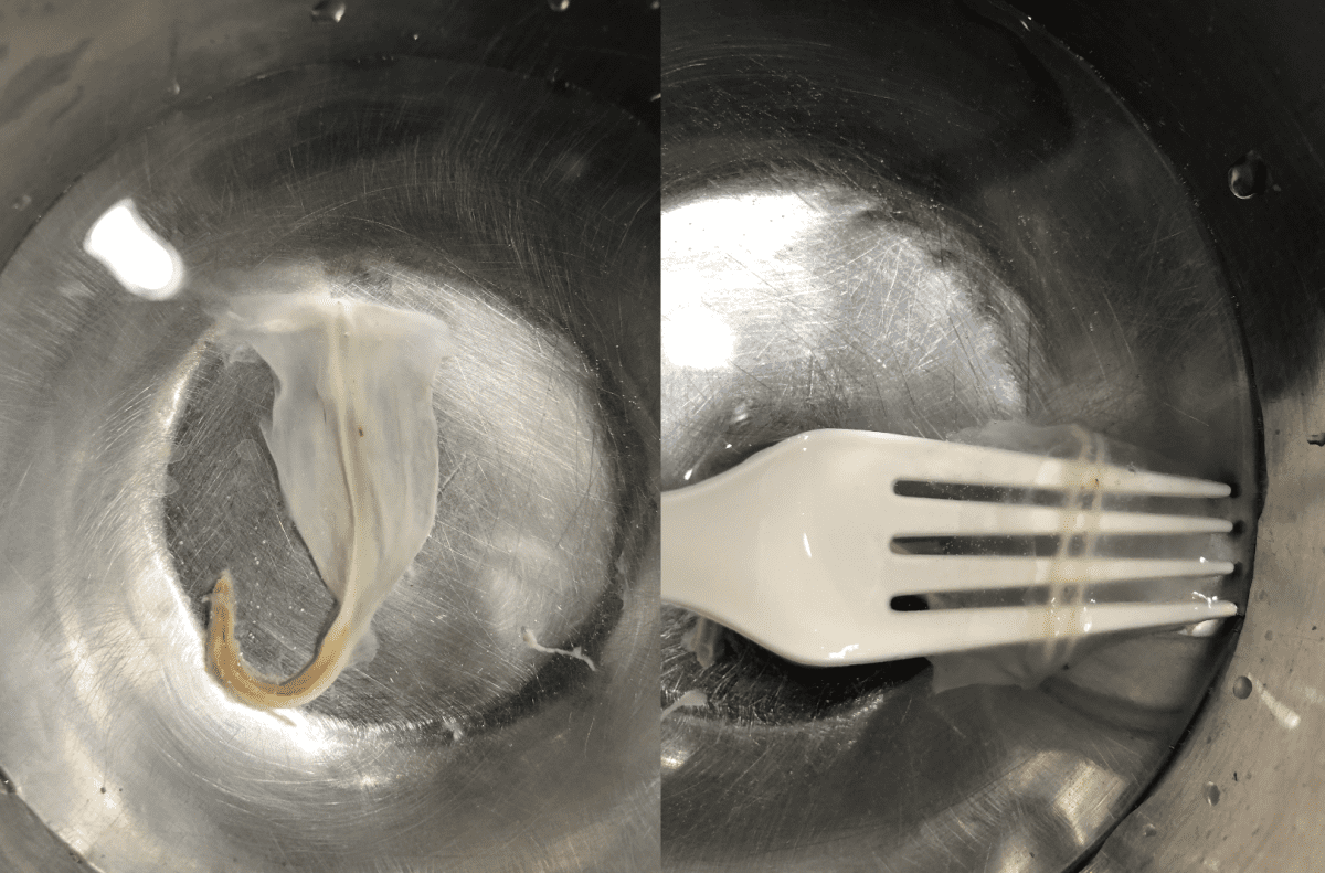
Suspected fluke images
1. A single dose of a parasite drug that treats flukes was taken before bed and the next morning this parasite was passed. No enema was done.
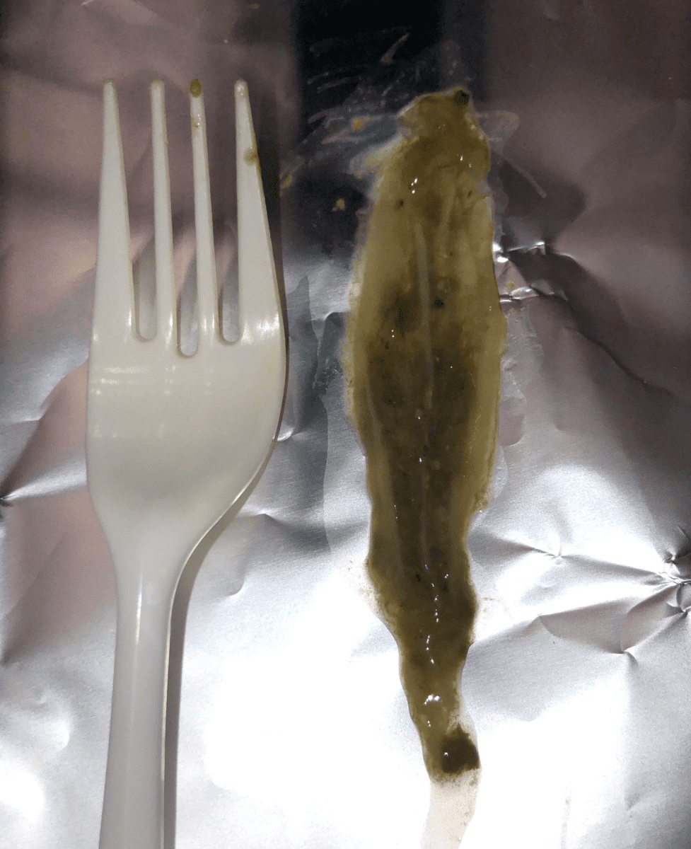
2. A single dose of a parasite drug was taken followed by two days of doing an oxidizing enema once a day, then this parasite was passed.
3 & 4. The following two parasite images were passed on different days while taking two parasite drugs that treat flukes plus oxidizing enemas were done once a day.
5. This parasite was passed after taking 2 parasite drugs that treat flukes and after an oxidizing enema. The parasite often splits open from being partially digested has it remains in the body for about 24 hours after the parasite is killed by the oxidizing agent. But the 2 characteristic veins or intestine structures are still visible.
Parasites are the main cause of all chronic disease. We have supported hundreds of students in many countries and we didn’t realize how common flukes were until our students started to submit parasite pictures that are very characteristic of flukes. These students also tested well for parasite drugs that treat flukes and as these parasites are passed, inflammation decreases dramatically and quickly.
It’s important to note that when an oxidizing enema is performed one morning and then a follow up oxidizing enema is done the next morning, the parasites that were killed by the the first enema will be passed with the second enema 24 hours later and thus will not be fully intact. As soon as the parasite dies, the body will start to break it down (digest it) so when it is released during the second enema, it will look partially digested. This is why the images of the parasites are not fully intact when students use oxidizing enemas.
It is our hope that this information helps you get to the root cause of the symptoms and disease that you may be dealing with and help you appreciate that intestinal fluke infections are much more common than we are told, even in developed countries.
There are real solutions to recover from parasites today!
To restore health, we must focus on treating the cause of inflammation, which are parasites. First, identify the enemy (parasites), then support the body and treat the parasites while following a holistic approach. When parasitic infections are treated effectively, we can overcome inflammation or disease.
If you’re frustrated with the fact that our standard of care STILL doesn’t offer a real solution for treating MS and other diseases, then click on the link below to watch Pam Bartha’s free masterclass training and discover REAL solutions that have allowed Pam and many others to live free from MS and other diseases.
CLICK Here to watch Pam’s masterclass training
References:
[i] https://www.uptodate.com/contents/intestinal-flukes
[ii] https://www.sciencedirect.com/topics/immunology-and-microbiology/fasciolopsis-buski
[iii] https://www.cdc.gov/parasites/fasciolopsis/biology.html#:~:text=After%20ingestion%2C%20the%20metacercariae%20excyst,span%20of%20about%20one%20year.
[iv] https://reference.medscape.com/slideshow/intestinal-parasites-6010996#9
[v] https://www.cdc.gov/parasites/fasciolopsis/faqs.html
[vi] https://emedicine.medscape.com/article/219662-workup#c7
[vii] https://emedicine.medscape.com/article/219662-treatment
[viii] https://en.wikipedia.org/wiki/Fasciolopsiasis#cite_note-2

Clinically diagnosed with multiple sclerosis at the age of 28, Pam chose an alternative approach to recovery. Now decades later and still symptom free, she coaches others on how to treat the root cause of chronic disease, using a holistic approach. She can teach you how, too.
Pam is the author of Become a Wellness Champion and founder of Live Disease Free. She is a wellness expert, coach and speaker.
The Live Disease Free Academy has helped hundreds of Wellness Champions in over 15 countries take charge of their health and experience profound improvements in their life.